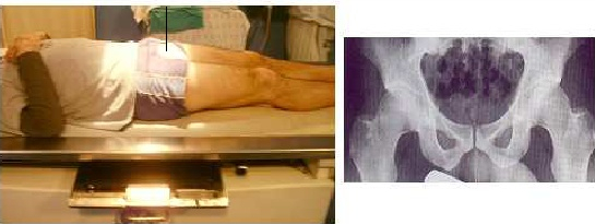
AP HIP PROJECTION
Anteroposterior • Unilateral or Bilateral View • Coxofemoral Joint Evaluation
Exposure Factors
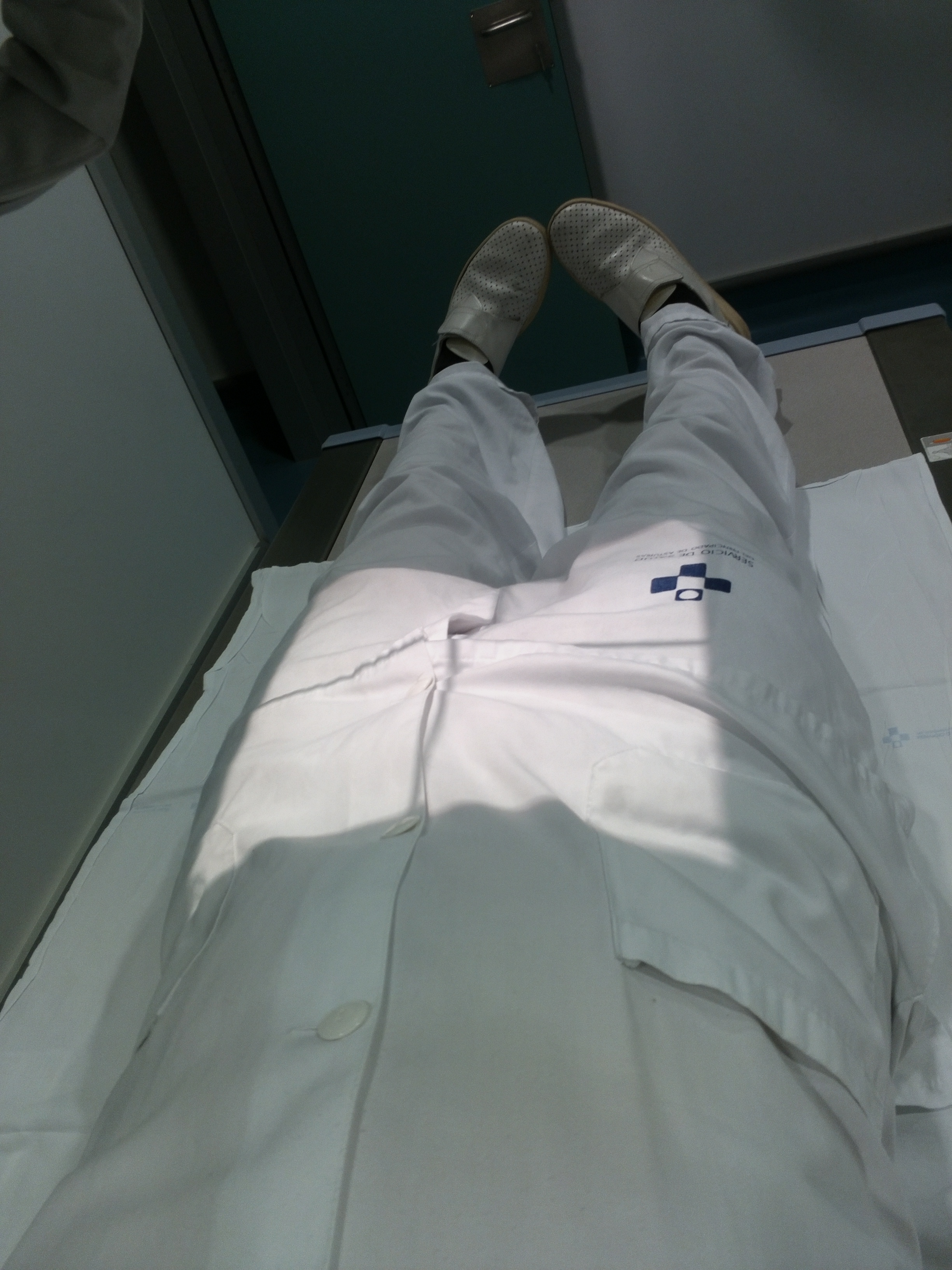
Equipment: With bucky. Position: Supine.
Plate Size
Visible Anatomical Structures
Coxofemoral Joints
One or both hips
Proximal Femurs
Upper portion
Trochanters
Greater and lesser
Coccyx
Caudal end
Sacrum
Distal portion
- Femoral neck - Portion between head and diaphysis
- Femoral head - Articulation with acetabulum
- Acetabulum - Cotyloid cavity
- Articular cartilage - Joint space
- Proximal part of femur - Upper diaphysis
Patient Positioning
Central Ray Direction
Vertical and perpendicular to center of line between trochanters
Bilateral study: Center of imaginary line between greater trochanters
Unilateral study: Center at femoral neck of affected hip
Angulation: 0° - Straight vertical
Patient Instructions
"Do not breathe during exposure"
Maintain complete immobility - Do not move legs during exposure
IMPORTANT NOTE
Unilateral study: When a single hip is requested, the central ray will be centered specifically on the femoral neck of the affected hip.
Patients with prosthesis: In patients with hip prosthesis, ensure the prosthesis appears complete in the image, including all its components.
Variations: Plate size and centering vary depending on whether the study is unilateral or bilateral comparative.
Technical Considerations
Selective Centering
Different centering depending on unilateral or bilateral study.
Prosthesis
Ensure complete visualization of all prosthetic components.
Plate Size
Adequate selection according to clinical need (unilateral/bilateral).
Clinical Indications
Unilateral vs Bilateral
UNILATERAL STUDY
Plate: 24 × 30 cm
Centering: Femoral neck
Indication: Specific pathology of one hip
BILATERAL STUDY
Plate: 35 × 43 cm
Centering: Line between trochanters
Indication: Comparison or general evaluation